


 Operating Microscope
MODERN OPERATIVE TECHNOLOGIES
Diagnostic imaging of brain tumors has been dramatically modified in the past three decades by the introduction of computer-assisted image reconstruction and advancements in basic sciences. MRI and CT scan have had a major impact on early diagnosis of brain tumors, thus permitting to operate patients in rather good neurological conditions, with better results in terms of quality of life and duration of survival.
At the same time, new operative tools have been introduced in clinical practice. These tools have deeply changed the surgery of brain tumors, with dramatic improvements of operative results and lessening of mortality and morbidity rates.
The neurosurgical operating room of the "Regina Elena" National Cancer Institute, Rome, Italy, has the most modern technologies.This is the most important one. Its daily use for 35 years has changed two generations of neurosurgeons. It permits to work under magnification and with an important illumination. Magnification ranges from 2 to 8 times. The most current operating microscopes are combined with a neuronavigator and partly robotized.
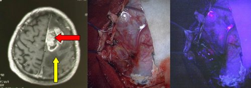
Fig.6.1: on the left, axial MRI of a left temporal glioblastoma. In the middle, operative appearance of the tumor; on the right, operative view with a fluorescent substance that marks the tumoral areas in red (arrows).
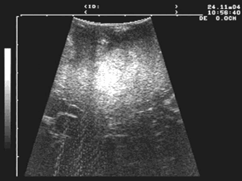
Video 6.1: This short video well shows the efficacy of intra-operative echography. See also Fig. 5.51.
Page 25
 Previous Page
